 Identificaciones de peptolab

Fotos / Sonidos

Qué
Género ColpodaObserv.
crseaquistDescripción
Gathered dry leaves on 2024-02-23 and stored in water.
 Picturepest, algunos derechos reservados (CC BY)")
Fotos / Sonidos

Qué
Nassula ornataObserv.
plingfactoryDescripción
very colourful ciliate due to digestion of cyanobacteria. More on this here:
https://www.plingfactory.de/Science/Atlas/KennkartenProtista/01e-protista/e-Ciliata/e-source/Nassula%20ornata.html
 Deuterostome, algunos derechos reservados (CC BY-SA)")
Fotos / Sonidos

Observ.
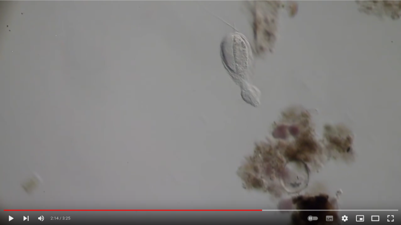
closterium_mysteriumDescripción
A water sample was taken from a small puddle in the playground. The sample had been in room temperature storage until assayed after 2 days.
Video: https://youtu.be/hnVz2mbtl7M
 Picturepest, algunos derechos reservados (CC BY)")
Fotos / Sonidos

Qué
Género ThuricolaObserv.
mnold1Descripción
Mag. 200x (1,4); 400X (2,3)
- A pond-side water sample (retentate) was taken on 01/06/2024 using a 10µ dip net to enrich for microorganisms. (The shoreline had a layer of ice about 1/4" thick that needed to be breached in order to accommodate the dipnet. The retentate contained a lot of sediment due to the shallow depth and disturbance caused by the net.) Air temp 35F.
Thuricola, a loricate peritrich ciliate. Lorica has a valve flap that must be pushed open to allow the zooids to feed. The lorica is round-bottomed and stalkless. Images in the video, https://youtu.be/om-CaI8zPlk, suggest there may be a vertical indentation at the aperture of the lorica.
 Giuseppe Vago, algunos derechos reservados (CC BY)")
Fotos / Sonidos

Observ.
closterium_mysteriumDescripción
This is one of three related observations of ciliates: two Stentor and its parasites Sphaerophrya from one water sample. This is an observation of parasitic ciliates (at different stages).
A water sample was taken from the bank of the Vuoksi River. The sample was stored at room temperature and observed one day after collection.
Related Observations:
1st Stentor https://www.inaturalist.org/observations/195742375
2nd Stentor https://www.inaturalist.org/observations/195742590
Video: https://youtu.be/rWhk8K2WvFE
 Илья Монашев, todos los derechos reservados, subido por Илья Монашев")
Fotos / Sonidos

Observ.
mhinczDescripción
Found in freshwater sample (edge of pond), 400x magnification.
 peptolab, algunos derechos reservados (CC BY-NC)")
Fotos / Sonidos

Observ.
dloarieDescripción
30 µ long. Apical mouth with liplike oral cavity and long mouth cilia. Long caudal cilium. Found on tree moss.
Phase contrast.
 peptolab, algunos derechos reservados (CC BY-NC)")

Fotos / Sonidos

Observ.
crseaquistDescripción
Water sample taken from the edge of a stagnate freshwater playa on 2023-11-06 using a turkey baster.
 zookanthos, algunos derechos reservados (CC BY), subido por zookanthos")

Fotos / Sonidos

Observ.
dms1535Descripción
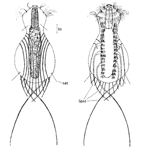
Haltidytes crassus was a relatively common species at the time of its first description. Since then - probably due to the influence of industrial agriculture - this pretty gastrotrich has become very rare.
The main characteristic of H. crassus are the five dorsal and three ventral “jumping spines”, with which the animal catapults itself out of the danger zone when threatened.
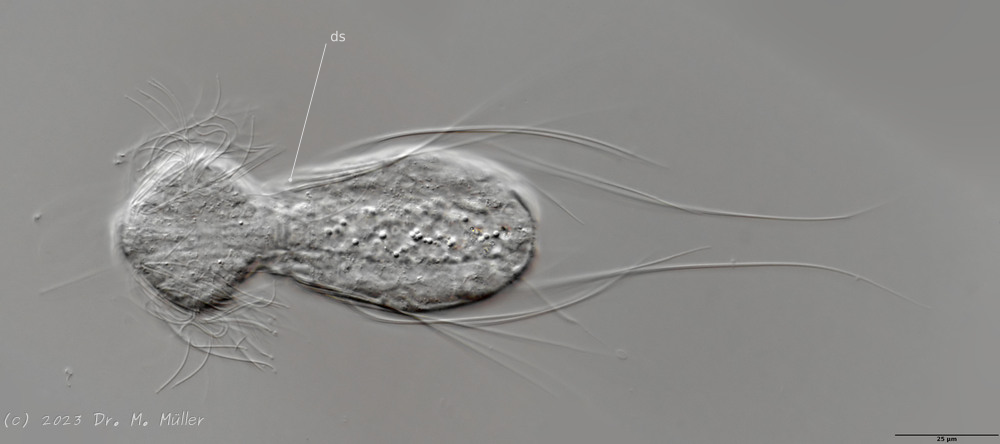 Dorsal (juvenile animal); ds: dorsal spines (5 pairs).
The long, caudal and hair-shaped last ventral pair of spines, which far overhang the body and cross each other in resting position, is conspicuous. Three ciliary rings on the head are used for swimming locomotion, while the ventral ciliary bands typical of gastrotrichs are used for gliding over the substrate.
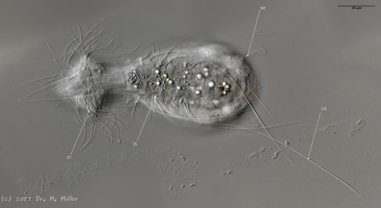
Ventral; cr: 3 ciliary rings for locomotion; js: jump spines; cs: long caudal spines; se: seta on papillae.
The posterior palps - as in all members of the genus Hatidytes - are on distinct papillae.
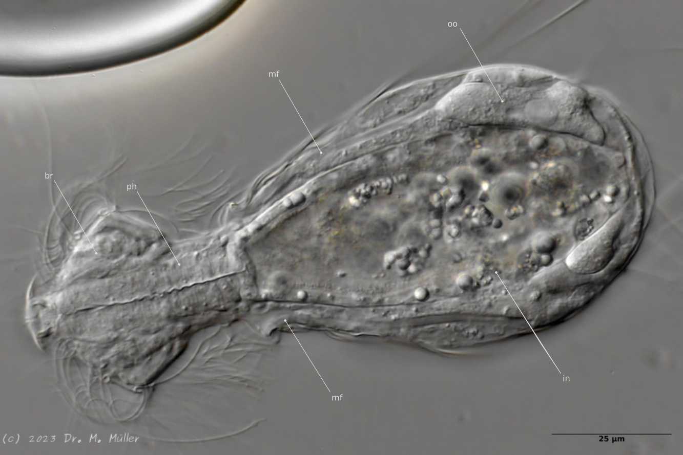
Median; br: “brain”; ph: pharynx; oo: mature ovum; in: “intestine”; mf: muscle fibers that move the jumping spines.
The internal anatomy has some peculiarities. For example, distinct muscle fibers can be seen that couple to the attachment sites of the jumping spines. Adhesive glands and the associated adhesive tubes are not present in this genus.
The typical movement and behavior of the animals can be seen in the following YouTube video:
Movie about Haltidytes crassus
 peptolab, algunos derechos reservados (CC BY-NC)")
Fotos / Sonidos

Observ.
mnold1Descripción
Mag. 400x
- A water sample was taken on 09/03/2023 from pond-edge using a 10µ dip net to enrich for microbes. Air temp. 80°F.
Short-spined Trachelomonas. Perhaps T. hispida as seen here in Fig. 42 https://www.researchgate.net/publication/314542785_Pigmented_Euglenophytes_in_a_natural_and_shallow_lake_in_the_semiarid_region_of_Paraiba_State_Brazil
 peptolab, algunos derechos reservados (CC BY-NC)")

 Lane Allen, algunos derechos reservados (CC BY-NC), subido por Lane Allen")


Fotos / Sonidos

Observ.
shanesmicroscopeDescripción
Continued observation of a sample collected 20210314 from a river tributary, scoop of sediment near the bank.
 zookanthos, algunos derechos reservados (CC BY), subido por zookanthos")
Fotos / Sonidos

Observ.
crseaquistDescripción
Water sample (freshwater) was taken on 09/09/2022 using a 10µm dip net to enrich for microorganisms.
Fotos / Sonidos

Observ.
crseaquistDescripción
Water sample (freshwater) was taken on 2023-04-09 using a turkey baster.
Fotos / Sonidos

Observ.
closterium_mysteriumDescripción
Here, a population of Cinetochilum margaritaceum is presented and measurements of several of them have been taken.
A water sample was collected from the shore of Srednerogatsky Pond. The air temperature was 14°C (57.2 °F). The sample was kept at room temperature until it was assayed 6 days later.
Video: https://youtu.be/yCe9MKw1KWQ

 Vicente Franch Meneu, algunos derechos reservados (CC BY-NC), subido por Vicente Franch Meneu")
Fotos / Sonidos

Qué
Bresslaua voraxObserv.
closterium_mysteriumDescripción
A predatory ciliate of the family Colpodidae that feeds on the ciliates Colpoda sp.
A sample was taken from the moss on the tree bark. The sample was stored at room temperature and observed one day after collection.
Video: https://youtu.be/S5B7_vhAbJQ
 peptolab, algunos derechos reservados (CC BY-NC)")
Fotos / Sonidos

Observ.
bdstaylorDescripción
Likely a Leptopharynx costatus with zoochlorellae.
Like other microthoracids, it is equipped with extrusomes. When exploded, these have four arms at the tip (a character of the family Microthoracidae, per Omar & Foissner, 2012, link below). The last two images show discharged extrusomes.
I looked at two specimens closely (the first is shown in images 1-3, the second in images 4 and 5). Both were a bit over 50 μm in length, and resemble those of L. bromeliophilus (as recorded in Omar & Foissner, 2011, link below).
 Igor Shelpiakov, algunos derechos reservados (CC BY), subido por Igor Shelpiakov")

 peptolab, algunos derechos reservados (CC BY-NC)")
Fotos / Sonidos

Qué
Ciliado (Halteria grandinella)Observ.
closterium_mysteriumDescripción
A water sample was taken from the shore of Srednerogatsky Pond. The air temperature was 14°C (57.2 °F). The sample was stored at room temperature and observed 10 days after collection.
Video: https://youtu.be/7Awc1MaxZm0
 peptolab, algunos derechos reservados (CC BY-NC)")
Fotos / Sonidos
Qué
Ileonema disparObserv.
bdstaylorDescripción
A ciliate with a curious flagellum-like process at its anterior. This structure is not a true flagellum. It does not move independently, but simply dangles from the ciliate's mouth as it swims around. Its purpose is not known. In other respects, Ileonema closely resembles Trachelophyllum. Like members of that genus it is enveloped by a coat of ornate epicortical scales, visible only in the scanning electron microscope. (See Foissner's monograph on Terrestrial and Semiterrestrial Ciliates from Venezuela and Galápagos, pp. 50ff)
Ileonema dispar is the type species of the genus, which was erected in 1884 by Alfred C. Stokes. In 1922, Penard described a similar species under the name I. simplex. I think there reason to believe that I. dispar and I. simplex are the same species, viewed in different conditions. Penard observed only one specimen, although he was able to watch it for a couple of hours. Stokes found I. dispar "in abundance", and describes it as having a variable shape: "Its changes of form are...quite constant as it rests among the algal threads and lengthens the entire body into a club-shaped creature searching for food, or contracts into an egg-shape with a short, narrow neck." However, he only illustrates one specimen (a rather pudgy one!), and it is this illustration that was reproduced in Kahl, with a short description (Kahl does not seem to have found the organism himself). Stokes also observed that his Ileonema is nearly identical in overall appearance to Trachelophyllum apiculatum: a telling comment, since the latter is usually quite slender, like Penard's simplex.
Stokes describes the "flagellum" as follows: "entire flagellum one-half the length of the body, the basal half thick, obliquely grooved and presenting a twisted or cord-like appearance, slightly tapering yet suddenly constricted at the beginning of the finely filamentous distal one-half."
The "flagella" of my critters are generally shaped much as Stokes describes: quite thick at the proximal end, finely filamentous at the distal. Seeing them at low magnification, it's easy to see how he might have misinterpreted the flagellum as being twisted, or ropelike. In my observations, the flagella of some individuals, at certain levels of illumination, appear to be quite uniform from base to tip.
The Ileonema in my population are also extremely contractile, ranging in shape from plumply ovoid to gracefully attenuated and vermiform. After they've eaten, they become almost unrecognizable!
 Bruce Taylor, algunos derechos reservados (CC BY), subido por Bruce Taylor")
Fotos / Sonidos

Qué
Ileonema disparObserv.
bdstaylorDescripción
A few more specimens of Ileonema dispar, the type species of the genus, which, as far as I know, has not been recorded since it was discovered by Stokes in 1884. It is differentiated from I. simplex by its flagellar process, which, in the species Stokes describes, is in two distinct parts, thick at the base and finely filamentous at the distal end (Stokes, 1884; Penard, 1922; Kahl, 1930). This is reflected in the name of the species, "dispar" (=unequal). Stokes also describes the species as having two contractile vacuoles in the posterior, an arrangement that would be extremely odd for a trachelophyllid ciliate. In several specimens, I observed some compartmentalization in the posterior vesicle, which could easily be read as two separate vacuoles adjacent to the "anal pore." See image #4.
The flagellar process is retractable, and its appearance is quite variable. The division of this organelle into two distinct parts--which I did not see in every specimen--strikes me as a weak character. As I noted in my previous observation, I think there is some reason to think that I. simplex should be considered a junior synonym of I. dispar (or possibly a variant, or sub-species).
I caught one in fission, and have included two images of the dividers.
I also watched an individual eating a huge clump of debris containing living and dead algae. This observation, and the contents of the cytoplasm in other individuals, lead me to think the critter lives by consuming small round green algae.
Fotos / Sonidos
Qué
Fabrea salinaObserv.
troy67Descripción
From an ultra-saline salt flat. Video is here: https://youtu.be/FqojSYv8tVU
 Troy Alexander, todos los derechos reservados, subido por Troy Alexander")
Fotos / Sonidos

Qué
Fabrea salinaObserv.
troy67Descripción
Darkly pigmented, oddly shaped ciliate common in this pool where mangroves were being propagated. High salinity - saltier than the nearby ocean.
Video uploaded here: https://youtu.be/NzBPgV_8RVk
Fotos / Sonidos
Qué
Fabrea salinaObserv.
troy67Descripción
Darkly pigmented, oddly shaped ciliate common in this pool where mangroves were being propagated. High salinity - saltier than the nearby ocean.
Video uploaded here: https://youtu.be/NzBPgV_8RVk
Fotos / Sonidos
Qué
Fabrea salinaObserv.
troy67Descripción
Darkly pigmented, oddly shaped ciliate common in this pool where mangroves were being propagated. High salinity - saltier than the nearby ocean.
Video uploaded here: https://youtu.be/NzBPgV_8RVk
Fotos / Sonidos
Qué
Fabrea salinaObserv.
troy67Descripción
Darkly pigmented, oddly shaped ciliate common in this pool where mangroves were being propagated. High salinity - saltier than the nearby ocean.
Video uploaded here: https://youtu.be/NzBPgV_8RVk
Fotos / Sonidos

Observ.
mnold1Descripción
Mag. 400x
Euglenoid with grit/sand-covered body, and a cytostome surrounded by a flared collar with a distinct rim. It uses a single flagellum for locomotion. It feeds on detritus and small algae. In the linked video you will see the creature worm its way through a mound of detritus, https://youtu.be/PUfNi0aoE5U. There were instances when it appeared to be foraging/feeding; the flared anterior moving over a surface like a handheld vacuum. :o) A very similar creature (but only in swimming rather than feeding mode) was observed from a different, local water-body https://www.inaturalist.org/observations/107162369.
- A water sample was taken on 5/8/2023, from the shore of Lantern Hill Pond, using a 10µ dip net to enrich for microbes. Air temp. 73°F.
 Nick Belliveau, algunos derechos reservados (CC BY-NC), subido por Nick Belliveau")
Estadísticas
- 60


